


Clinical Case I
A complex and comprehensive case that I treated start to finish as a student dentist.
About the Patient: 42-year old, female

Full Mouth Series
Findings:
Root tips #s: 4, 5, 12, 13, & 18 → EXT
Fractured cusps #s: 2 & 14 → EXT
Recurrent caries #s: 15 & 19 → B/U + Crown
Incipient lesion #: 29-D
Carious lesions: #s: 3-OL & 31-MO

Panoramic Radiograph
NSF.

Intraoral Photos
Noted with significant findings
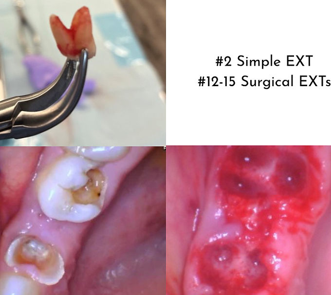
Extractions
Both simple and surgical extractions were completed on this patient. Pre-authorizations were submitted for the surgical extractions. Sutures were placed over the ridges of #12 -14 using the figure 8 method.

Resin Infiltration Treatment Icon)
Icon is an enamel remineralization treatment completed in one appointment to treat white spot lesions and incipient caries These photos were taken 4 months apart.

Removable Partial Denture (RPD)
Though not completed yet, this is the design I plan to use for the RPD.